The circulatory system, also known as the cardiovascular system, is made up of the blood, heart and blood vessels. Blood circulates in the system using two routes: systemic circulation and pulmonary circulation. The heart is part of both pathways.
The main structures of the circulatory system are identified in the following image
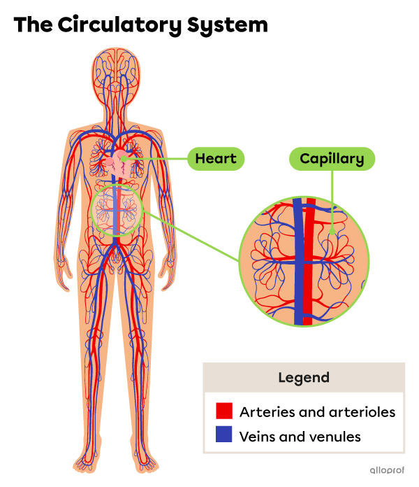
The circulatory system plays the following roles.
-
Transporting respiratory gases, nutrients and waste throughout the body.
-
Participating in the exchanges of gases, nutrients and waste between the cells and bloodstream.
The heart, its chambers and blood vessels are identified and described in the image and in the following table.
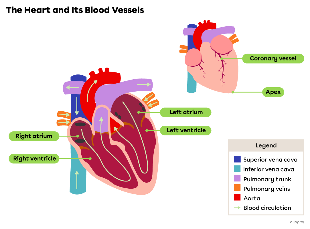
Note: As the heart is shown from the front, the right side and the left side are inverted in the image.
| Description | Functions |
|---|---|
|
The heart is a hollow muscular organ that acts like a double pump separated vertically by a partition, called a septum. The muscle tissue of the heart is also called myocardium. The heart is triangular in shape and its tip, called the apex, points slightly to the left and forward. |
|
|
The atria are the two upper chambers of the heart, referred to as the right atrium and the left atrium. The walls of the atria are made of cardiac muscle tissue, which is much thinner than the walls of the ventricles. |
Right atrium:
Left atrium:
|
|
The ventricles are the two lower chambers of the heart, referred to as the right ventricle and the left ventricle. The thick muscular tissue of their walls enables the ventricles to undergo powerful contractions when they are filled with blood. The wall of the left ventricle is also thicker than the wall of the right ventricle. |
Right ventricle:
|
| Description | Functions |
|---|---|
|
The coronary vessels are small blood vessels on the external surface of the heart. |
Carry blood rich in |\text{O}_2| to the cardiac muscle, then blood rich in |\text{CO}_2| to the right atrium. |
|
The superior vena cava and the inferior vena cava are veins. They both lead to the right atrium as part of the systemic circulation. |
Superior vena cava: Inferior vena cava: |
|
The pulmonary veins include two right pulmonary veins (coming from the right lung) and two left pulmonary veins (coming from the left lung). Together, the four pulmonary veins carry blood to the left atrium as part of the pulmonary circulation. |
Carry blood from the lungs to the left atrium. |
|
The pulmonary trunk is an artery. It splits into two smaller arteries, called the pulmonary arteries, that each go to one of the two lungs. The pulmonary trunk is part of the pulmonary circulation. |
Carries blood from the right ventricle to the lungs. |
|
The aorta is an artery. Exiting the left ventricle, the aorta goes over the pulmonary trunk, then splits into several smaller arteries. Some of them carry blood to the head and arms, while others carry blood to the abdomen and legs. The aorta is part of the systemic circulation. |
Carries blood from the left ventricle to the organs of the upper and lower body. |
All blood vessels form a one-way circuit through which blood is pumped when the heart contracts. Arteries, arterioles, capillaries, venules and veins are different types of blood vessels. Their structure (lumen diameter, thickness, elasticity, etc.) and their tissues are adapted to their specific role in the circulatory system.
-
Blood vessels are tubes that carry blood.
-
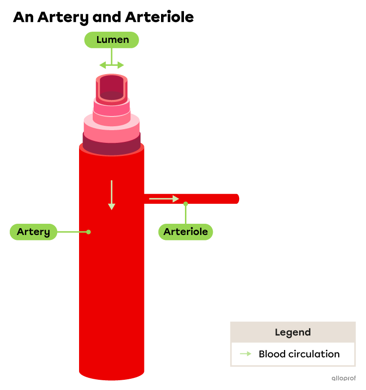
The lumen of a blood vessel is the empty space inside it where blood circulates.
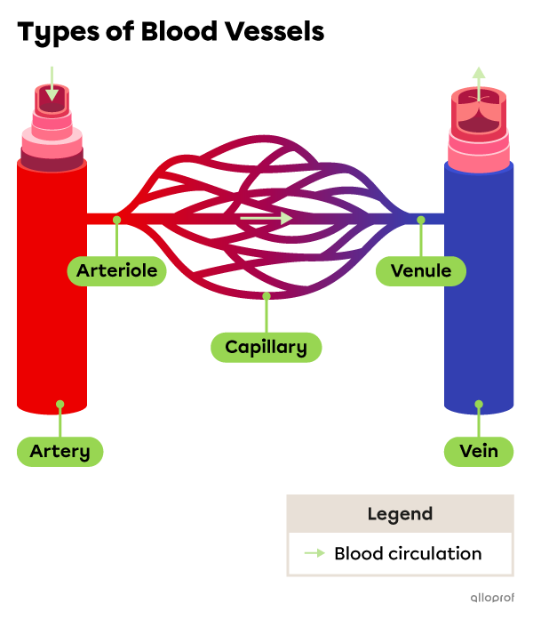
Arteries are blood vessels that carry blood from the heart to the organs.
On diagrams, arteries are often shown in red, but in reality they are colourless or slightly yellowish.
| Description | Function |
|---|---|
|
An artery is a blood vessel with a thicker and more elastic wall than the wall of a vein. In systemic circulation, blood carried by arteries, such as the aorta, is rich in oxygen |(\text{O}_2).| In pulmonary circulation, blood carried by arteries, such as the pulmonary trunk, is rich in carbon dioxide |(\text{CO}_2).| |
Carries blood from the heart to the arterioles, then to the capillaries of the organs. |
|
An arteriole is a small blood vessel that splits from an artery and branches off into several capillaries. |
Carries blood from an artery to several capillaries. |
-
The arteries experience higher blood pressure than veins because of blood pumped by the heart’s ventricles.
-
The wall of an artery is made up of layers of epithelial tissue, connective tissue, smooth muscle tissue and elastic fibres.
-
The diameter of an artery’s lumen varies depending on how close it is to the heart. Arteries near the heart generally have wider lumens.
| Description | Functions |
|---|---|
|
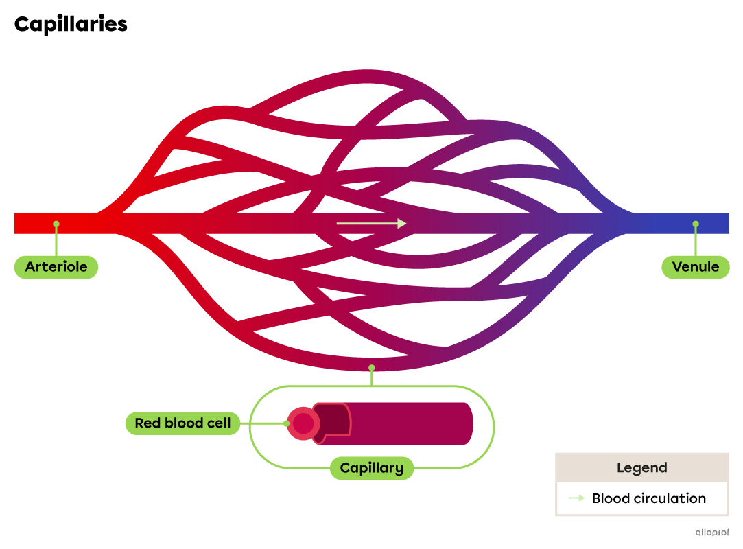
Capillaries are small vessels that connect the arterioles to the venules. They have thin, permeable walls. The lumen of a capillary is just wide enough to allow a single red blood cell through at a time. They have to circulate in a single file. |
|
Veins are blood vessels that carry blood from the organs to the heart.
On diagrams, veins are often shown in blue to easily differentiate between veins and arteries. This does not reflect the colour of blood. Blood in veins is not blue, it is generally dark red. It is dark red because of a low concentration of oxygen |(\text{O}_2).| Blood in pulmonary veins is bright red due to a high concentration of |\text{O}_2.|
When looking at wrist veins under the skin, they can appear blue and sometimes green. The reason for this is distorted perception of the colour resulting from the contrast with the colour of the skin. This effect is less obvious when the person’s skin is darker. In reality, regardless of the colour of the skin, veins are colourless or slightly yellowish.

sruilk, Shutterstock.com

Andrey_Popov, Shutterstock.com
| Description | Function |
|---|---|
|
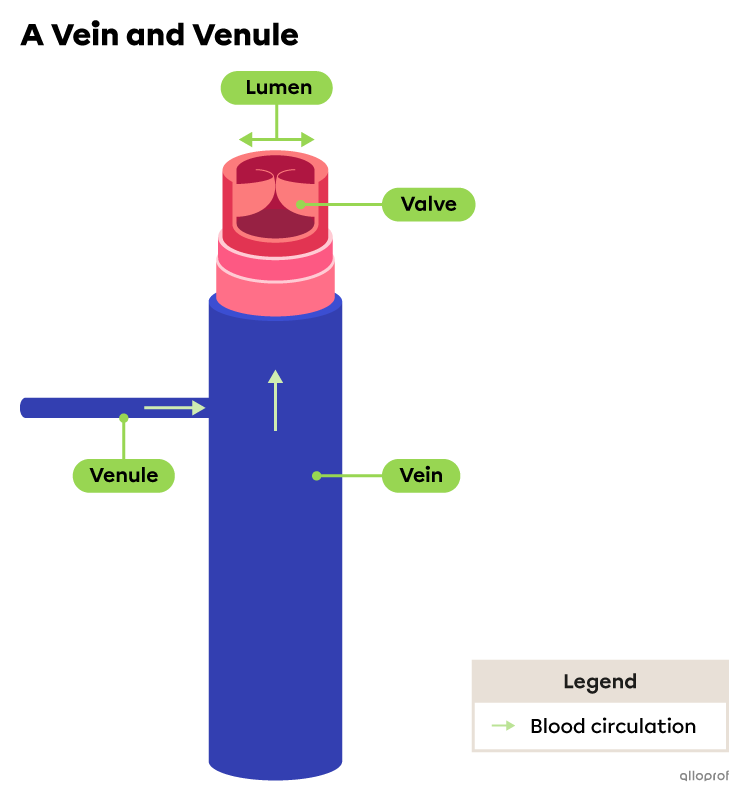
A venule is a small blood vessel formed where multiple capillaries join before connecting to a vein. |
Carries blood from the capillaries to the veins. |
|
Veins are blood vessels with a thinner and less elastic wall than arteries. Most veins have valves. They prevent venous reflux. In systemic circulation, blood carried by veins, such as the superior and inferior vena cava, is poor in oxygen |(\text{O}_2).| In pulmonary circulation, blood carried by veins, such as pulmonary veins, is rich in oxygen |\text{O}_2.| |
Carry blood from the systems and their organs to the heart. |
-
A valve is a small flap of tissue inside a vein. It allows blood to circulate in one direction and prevents blood from flowing backwards.
-
Venous reflux occurs when blood flows backwards through a vein rather than continuing towards the heart. Low blood pressure and gravity can cause venous reflux, mainly in the veins of the legs.
-
The blood pressure in veins is much lower than in arteries.
-
The wall of an artery is made up of layers of epithelial tissue, connective tissue, smooth muscle tissue and elastic fibres.
-
The diameter of the lumen of a vein varies depending on how close it is to the heart. Veins near the heart generally have wider lumens.