The material covered in this concept sheet goes beyond what is covered in high school. This is enrichment information for students who want to find out more.
The lymphatic vessels contain a fluid called lymph, which is composed of circulating interstitial fluid (extracellular fluid), as well as fluid from the liver and intestines. Lymphatic vessels are arranged so that lymph travels one way from the blood capillaries to the heart.
The main cells found in lymph are lymphocytes. While they are very common in the body, lymphatic vessels are not found in teeth, bone, marrow and myocardium. Lymphatic vessels in the central nervous system were discovered in 2015.
The lymphatic capillaries are the most upstream structures of the lymphatic network. Their extremities are blunt-ended. These capillaries are found between the cells and blood capillaries of the organism’s loose connective tissue. Their extremities are very permeable due to gaps in the membrane (much like roofing shingles installed upside down). This structural arrangement acts like a valve where the imbalance between the external and internal pressure enables fluids to flow from the outside to the inside. When the external pressure increases, the gaps open and will close when the internal pressure increases.
Lacteal vessels are highly specialized lymphatic capillaries found in the small intestine, more precisely in the villi of the mucosa. They carry a lymph called chyle from the small intestine. These vessels play an important function in the absorption of lipids.
The collecting vessels are downstream of the lymphatic capillaries, so they collect lymph from them. Their structure is quite similar to veins but they contain many more valves. In addition, their three membrane layers are much thinner and form many anastomoses, which are connections between vessels of the same type (a network).
While the superficial lymphatic vessels tend to run parallel to the veins, the deep lymphatic vessels follow the arteries, forming anastomosis around them.

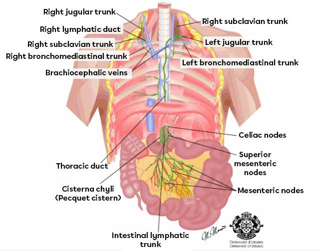
Lymphatic trunks are even larger in diameter than the lymphatic vessels and consist of collector lymphatic vessels that join into a larger one.
These trunks receive lymph from upstream vessels to drain large areas of the body. Most trunks come in pairs: lumbar, bronchomediastinal, subclavian and jugular. However, there is only one intestinal trunk. The names of the trunks are largely inspired by the region where the lymph comes from.

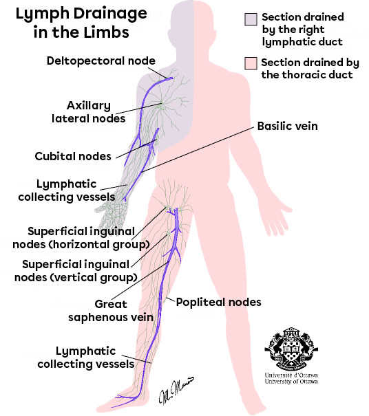
All circulating lymph ends up in two large ducts: the right lymphatic duct and the thoracic duct.
The right lymphatic duct receives lymph from the right arm, as well as from the right side of the head and thorax.
The thoracic duct receives lymph from the rest of the body and is therefore larger. It starts in front of the first lumbar vertebrae in the form of a sac, called the cisterna chyli, sometimes referred to as the Pecquet cistern. The lymph from the intestinal trunk and the two lumbar trunks (which drain the lymph of the lower limbs) is collected by the cisterna chyli. Lymph from the left side of the thorax and head, as well as the left arm, is collected as it travels from the thoracic duct to the upper part of the body. These two ducts then discharge lymph content into the venous system, each on its own side. In this case, the target is the subclavian vein, at the junction of the internal jugular vein.
The main and most well-known lymphatic organs are the lymphatic nodes, or lymph nodes.
They are distributed across the lymphatic system, but there are some clusters of nodes in the neck, groin, armpit and abdominal cavity. In these areas, they are large enough and close enough to the skin’s surface to be perceived by touch.
There are hundreds of lymph nodes, ranging in size from 1 to 25 mm. They resemble beans.
Other major lymphatic organs are the tonsils, spleen, thymus and lymphatic follicles.
Tonsils are located in the pharyngeal region and are generally the most structurally simple of the lymphatic organs. They often become infected and are often removed surgically.
The spleen is the largest lymphatic organ. It is highly vascularized and located to the left of the stomach.
The thymus is most active in the early years of life and becomes less active later on. It is located in the thorax, between the lungs, and partially covers the upper part of the heart.
Finally, clusters of lymphatic follicles, also known as Peyer’s patches, are located around the edge of the small intestine and have a structure similar to that of the tonsils. Another cluster is found in the wall of the appendix.
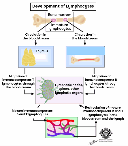
The primary function of lymph is immunity, which is why white blood cells that participate in the defence of the organism are called lymphocytes.
Upon entering the lymphatic system, any antigen (any particle foreign to the body such as bacteria, viruses, cancer cells, incompatible erythrocytes, etc.) will be attacked by T- or B lymphocytes, or by phagocytes (involved in phagocytosis).
In a lymph node, several types of cells combine to create a trap for foreign bodies resembling a spider’s web. The structural support of the node is provided by the reticular cells. The T lymphocytes and dendritic cells activate the inflammatory response and can directly attack and destroy foreign cells.
Lymphocytes also create plasma cells which secrete the antibodies necessary to inhibit the action of the antigens, until they are absorbed (phagocytosed) by macrophages.
The lymph serves as a development area for lymphocytes, providing them with shelter and a strategic position against attacks from infectious cells.

The second function is related to the drainage of interstitial fluid. This drainage prevents edema, which is the accumulation of fluid, limiting infections and swelling. It also provides a better distribution of interstitial fluids throughout the body. This allows for osmotic equilibrium between the tissues and blood.
The lymphatic system has no pump, unlike the arterial blood in the heart. The pressure is usually quite low, which limits the possibility of taking advantage of a strong pressure gradient.
The propagation mechanism used by the lymphatic system is very similar to that of venous return. First, there is a propulsion mechanism involving muscles, the skeleton, peristalsis and respiration. The various movements of the body compress and relax the lymphatic vessels, acting as a pump. This causes the lymph to change compartments, blocked off by valves that prevent reflux. Some of the pressure from the interstitial fluid is used to move the lymph forward. Lymphatic vessels attached to the arteries can be found in the same connective tissue layer. The pulsation in the arteries causes pressure changes resulting in a movement of the lymph. Finally, lymphatic ducts and lymphatic trunks can engage in rhythmic contractions that push lymph into the vessels, even though this mechanism is less important than the contractions caused by the musculoskeletal system.
The purpose of a lymph node, and what probably gave it its name, is to contain the lymph long enough to allow the lymphocytes to act.
There are several afferent (incoming) vessels but only one efferent (outgoing) vessel. The centre of the lymph node is a type of sinus where lymph is retained. It creates a circulation blockage, which causes lymph to stagnate in the lymph node. This gives lymphocytes time to purify part of the lymph from infectious agents. There are usually several neighbouring nodes in the same pathway, which allows the lymph to be fully purified by passing through several of these nodes.