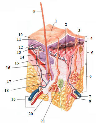
The skin is the organ that senses touch and perceives tactile, thermal and painful stimuli. It is the largest organ of the human body and accounts for about 7% of the body’s mass. The skin is composed of three layers: the epidermis, dermis and hypodermis.

- Pore
- Dermal-epidermal junction
- Nerve ending
- Epidermis
- Dermis
- Hypodermis
- Vein
- Artery
- Hair
- Stratum corneum (outermost layer of the epidermis)
- Pigmented layer (in the epidermis)
- Keratinocytes
- Melanocytes
- Hair erector muscle
- Sebaceous gland
- Hair follicle
- Bulb
- Nerve
- Lymphatic and blood vessels
- Sweat gland
- Pacinian corpuscle
The epidermis is made up of four distinct layers from the outside in: the stratum corneum, the stratum granulosum, the stratum spinosum and finally the stratum basale. The stratum corneum is the upper layer of the skin. It’s impermeable and varies in thickness on different areas of the body. This layer is made up of flattened dead cells that are constantly being replaced. The next layers contain two important cell types: keratinocytes and melanocytes. The keratinocytes produce keratin, a fibrous substance that gives the skin extra strength and protects it against wear and tear and pathogens. The melanocytes synthesize melanin, a pigment that protects the skin from the harmful effects of sunlight, in other words, these cells are responsible for tanning. The cell division necessary for skin renewal takes place in the stratum basale. The epidermis of the entire body is renewed on average every 60 days.
The dermis is located just under the epidermis and is a resistant and flexible tissue. It’s richly innervated and many lymphatic and blood vessels run through it. The hair follicles (hair roots), sebaceous glands (secrete an oily substance called sebum) and sweat glands (secrete sweat) are all located in the dermis. The sweat glands are connected to the surface by a duct that opens into a pore, which allows sweat to escape. The base of each hair has an erector muscle which is responsible for straightening the hair, which is known as goose bumps. Finally, the dermis contains numerous sensory receptors that receive different stimuli (pressure, temperature, pain) as well as nerve endings that transform these stimuli into nerve impulses.
The hypodermis is located under the dermis and is a barrier between the dermis and the rest of the body (especially muscles and tendons). It’s mainly made up of fatty tissue and serves, among other things, as an energy reserve and thermal insulator. The hypodermis doesn’t have the same thickness everywhere on the body: it’s thicker in certain places, for example on the stomach and buttocks.
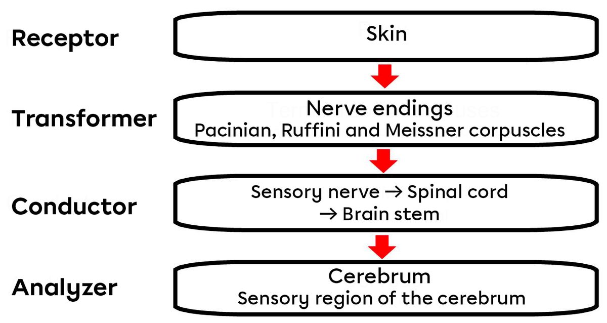
Stimuli are perceived by the skin through sensory receptors. The nerve endings, such as the Pacinian, Ruffini and Meissner corpuscles (all of which react to pressure), as well as free nerve endings (which react to temperature and pain), convert the stimuli into nerve impulses. The signal is then sent to the cerebrum via a sensory nerve. If the stimulus comes from below the head, the impulse travels through the spinal cord to the brain stem, before arriving at the sensory area of the cerebrum. The sensation caused by the original stimulus is perceived.

In addition to being the organ associated with touch, the skin also performs other functions such as protection, thermal regulation, absorption (for example, of cream or medication), excretion and production of vitamin D.
Protection is, of course, one of the skin’s most important functions. The surface layer of the skin (the stratum corneum) protects against bacteria. The sebum produced by the sebaceous glands makes the skin waterproof and the melanin protects it from the sun’s UV rays.
The skin’s protection function
The skin forms a resistant barrier to at least three types of potential hazards. First, it’s a physical or mechanical barrier. In fact, besides covering the entire body, it is resistant to abrasion thanks to the keratinized cells. The fact that the skin is continuous helps to protect the body against bacterial invasions. Water and water-soluble substances are also retained in the body because the skin does not allow their diffusion. All this provides a physical filter that prevents most substances from entering the body. A chemical barrier is provided by a film of acidic liquid contained in the skin’s secretions. Although the epidermis is covered with bacteria, this acidic film delays their multiplication.
The cells of the epidermis also secrete a natural antibiotic (human defensin) that pierces the bacterial walls. Cathelicidin is a protein that’s released at the site of a wound and prevents the invasion of type A streptococci. Melanin, on the other hand, is a pigment that protects DNA from UV rays that cause mutations and cancers. Finally, the biological barrier is provided by macrophagocytes in the dermis and epidermis and by the DNA itself. Macrophagocytes are able to eliminate most pathogens and recognize foreign antigens. The electrons of the DNA are able to absorb solar radiation (especially UV) and transmit this energy to the surrounding molecules. The energy is then absorbed and converted back into heat.
Regulating body temperature is the skin’s second function. When it’s hot, the dermal blood vessels dilate and the sweat glands secrete more sweat. At room temperature (between 20°C and 25°C), about 200 mL of sweat is produced per day. In an extreme heat situation, though, it’s possible to lose up to 1 L of sweat in one hour! Conversely, if it’s very cold, the capillaries of the dermis contract and the sweat glands won’t be stimulated. This contraction reduces the blood flow in the skin in order to prioritize the blood supply to the body’s internal organs. The hypodermis is a layer of adipose tissue that also helps maintain internal temperature by acting as a thermal insulator.
The skin also has the role of producing vitamin D. The cholesterol molecules circulating in the blood vessels of the epidermis are transformed into vitamin D by sunlight. This vitamin helps absorb the calcium and phosphorus necessary for the proper development of the body’s bone structure.
Finally, the skin also plays a role in excretion. A certain proportion of the nitrogenous waste produced by the body (such as urea) is excreted through sweat. Water and minerals are also excreted in large quantities with sweat.
The skin is also a blood reservoir. It’s richly vascularized and can contain up to 5% of the body’s blood volume. The vessels can then be constricted when a nearby organ or muscle requires more oxygen.