The digestive system is the set of organs involved in the transformation of food into nutrients that can be absorbed by the body. It also enables the elimination of solid waste.
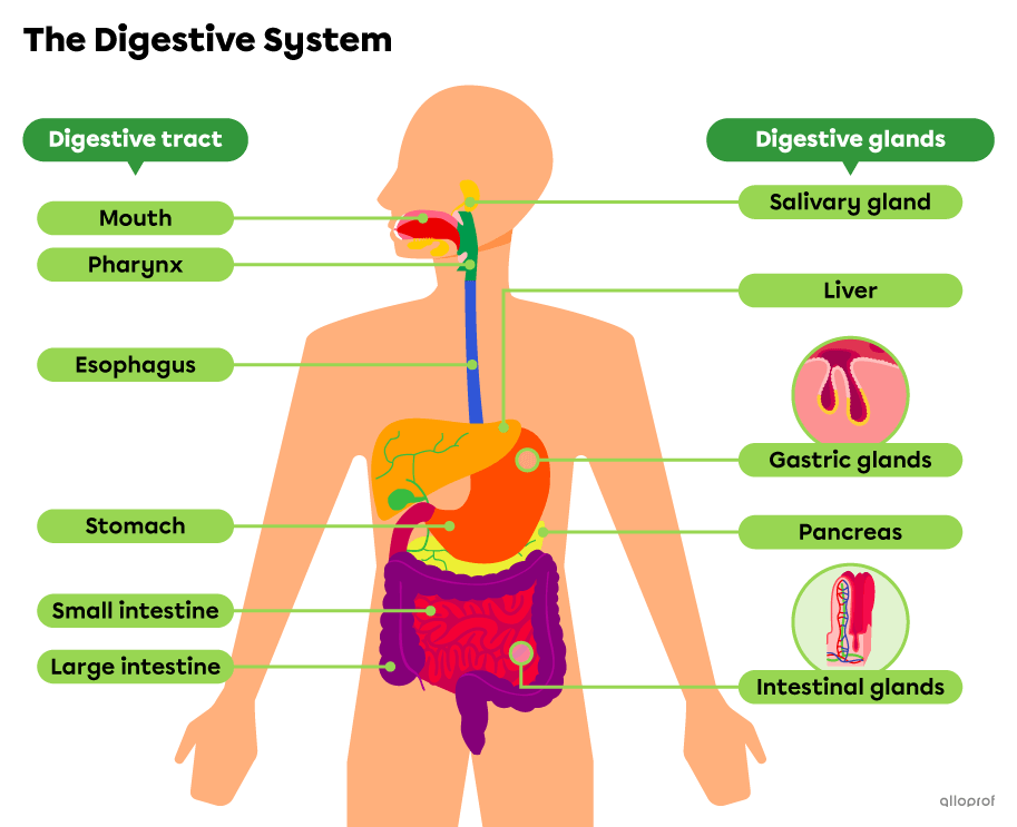
The digestive system consists of the digestive tract and the digestive glands.
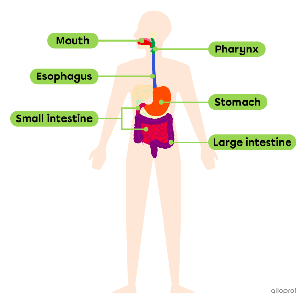
The digestive tract, also called the gastrointestinal tract or GI tract, is a long muscular tube through which the ingested food travels.
The role of the digestive tract is to break down food, absorb nutrients and eliminate waste.
The following image shows the organs of the digestive tract.
| Description | Functions |
|---|---|
|
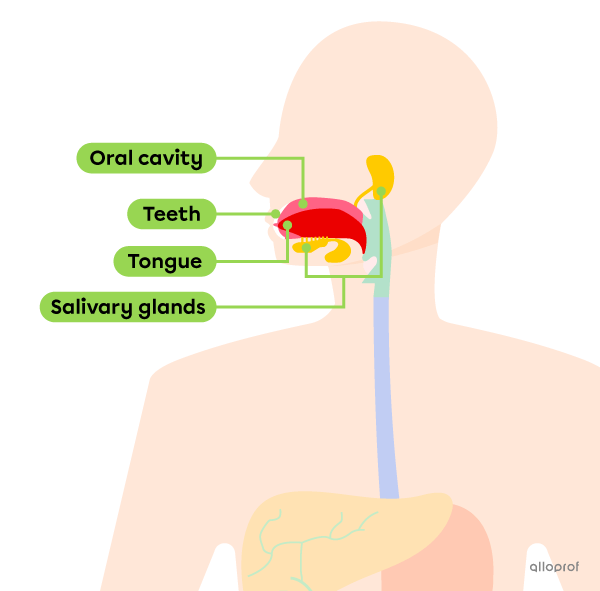
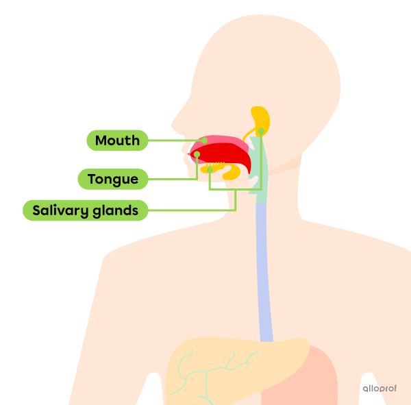
The mouth, or oral cavity, is a cavity lined with a mucous membrane, defined in front by the lips, on the sides by the cheeks, on top by the palate and in the back by the pharynx. The mouth includes certain accessory organs such as the teeth, tongue and salivary glands. |
|
| Description | Function |
|---|---|
|
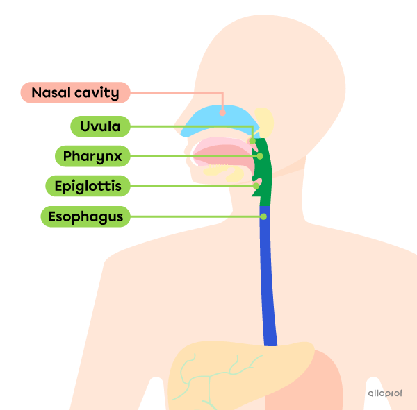
The pharynx is a muscular duct lined with a mucous membrane, connecting the nasal cavity, mouth and esophagus. It is a shared part of the respiratory and digestive systems. |
|
The upper part of the pharynx contains a muscle called the uvula. The lower part of the pharynx contains a cartilaginous flap called the epiglottis. These structures are involved in swallowing.
| Description | Function |
|---|---|
|
The esophagus is a tube about 25 cm long connecting the pharynx and the stomach. The walls of the esophagus contain smooth muscle tissue. |
|
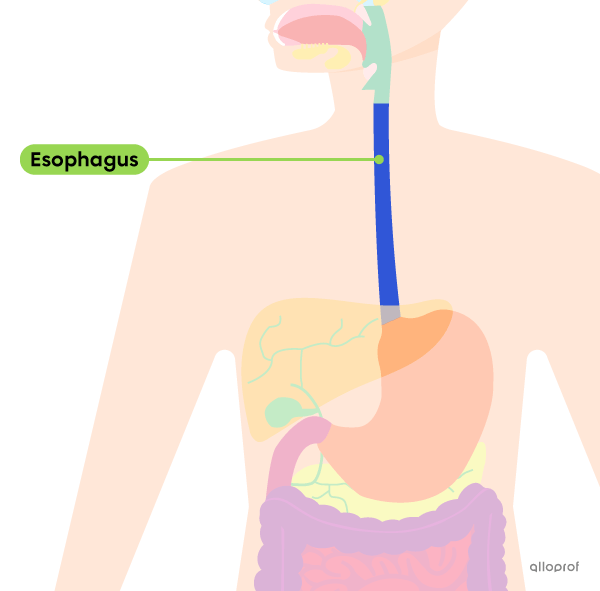
| Description | Functions |
|---|---|
|
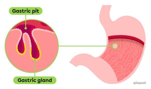
The stomach is a muscular pouch located between the esophagus and the small intestine. The epithelial tissue that lines the inside of the stomach is covered with microscopic cavities that contain the gastric glands. |
|
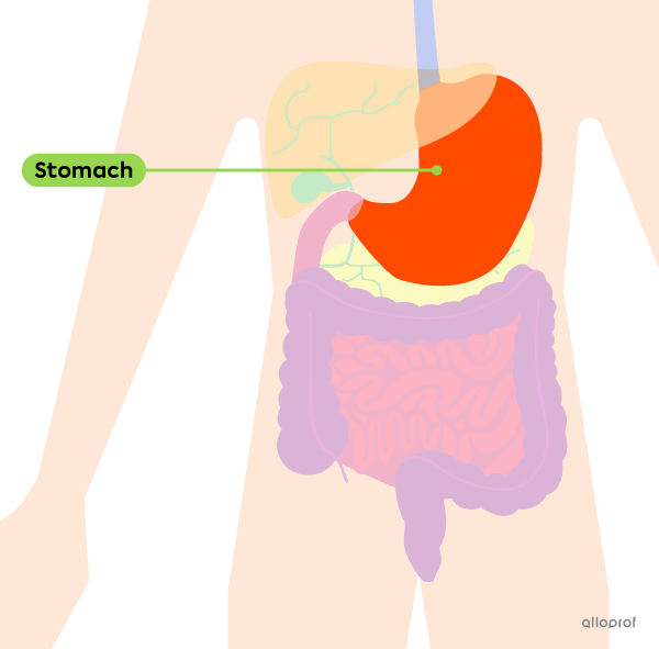
The elasticity of the stomach wall allows it to hold up to 4 L of food while its volume is only 50 mL when completely empty.
|
Description |
Functions |
|---|---|
|
The small intestine is a long muscular tube about 2.5 to 4 cm in diameter. It is located between the stomach and the large intestine. It is the longest organ in the digestive system. |
|
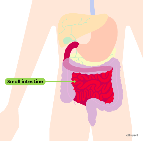
The epithelial tissue that lines the inside of the intestine forms thin, elongated, hair-like projections. These are the intestinal villi.
Each cell covering the surface of the intestinal villi is also covered with microscopic projections called microvilli.
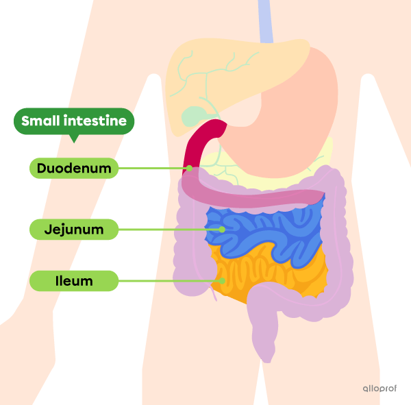
The small intestine is the longest organ of the digestive tract. While the total length of the digestive tract is about 9 m at rest, the small intestine is approximately 6-7 m long. It is subdivided into 3 sections: the duodenum, jejunum and ileum.
The duodenum is the shortest section of the small intestine. The stomach contents, bile and pancreatic juice are emptied into the duodenum. A significant part of chemical digestion occurs in this section.
Nutrients and some water are then absorbed in the jejunum and ileum.
| Description | Functions |
|---|---|
|
The large intestine is a muscular tube about 7 cm in diameter. The large intestine is the last organ of the digestive tract. |
|
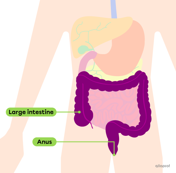
The terms colon and large intestine are not synonymous. In fact, the colon is only one section of the large intestine.
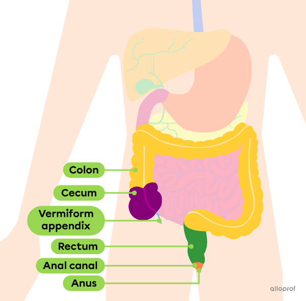
The large intestine is divided into several segments, including the cecum, vermiform appendix, colon, rectum and anal canal.
The cecum is the section of the large intestine that receives food waste from the small intestine.
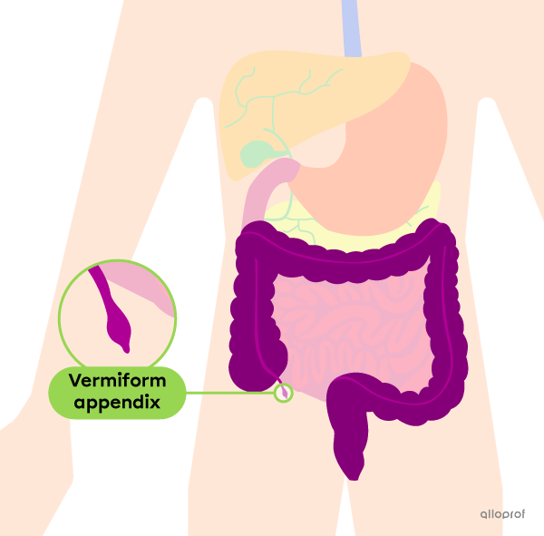
Connected to the cecum is a small extension of about 5-10 cm called the vermiform appendix, or simply appendix. This structure plays a role in the body's immune system.
The colon is the section between the cecum and the rectum. It is the longest segment of the large intestine. It helps absorb some of the remaining water and pushes food residue into the rectum.
The rectum is the section of the large intestine where fecal matter accumulates before defecation.
The anal canal is the last segment of the large intestine. It connects the rectum to the anus.
The anus is the opening that allows the elimination of feces from the body during defecation.
Appendicitis is the spontaneous inflammation of the vermiform appendix.
Because of its shape and position, sometimes the inside of the appendix becomes obstructed with feces, which can lead to infection. The infection can then cause the appendix to swell, which can damage the wall and eventually rupture. To avoid complications from a ruptured appendix, it is usually removed at the first sign of appendicitis.
Appendicitis is more common in adolescents, as the opening of the appendix is at its widest during this period of growth.
Digestive glands are glands that secrete substances that help break down food.
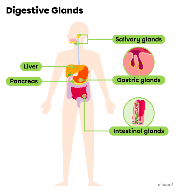
There are two types of digestive glands: integrated and accessory digestive glands.
|
Type of digestive glands |
Integrated digestive glands |
Accessory digestive glands |
|---|---|---|
|
Description |
These are digestive glands integrated directly into the wall of the digestive tract. They are in direct contact with the contents of the digestive tract. |
These are digestive glands located outside of the digestive tract. Their secretions are carried into the digestive tract through various ducts. |
|
Examples |
| Description | Function |
|---|---|
|
Salivary glands are accessory digestive glands located at the periphery of the mouth. They are mainly found under the tongue and at the back of the mouth. Other small salivary glands (not visible in the image) are integrated into the mucous membrane of the mouth. |
|
| Description | Function |
|---|---|
|
Gastric glands are digestive glands that are integrated into the inner wall of the stomach. They are located at the bottom of microscopic cavities, called gastric pits. |
|
| Description | Function |
|---|---|
|
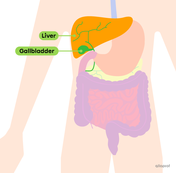
The liver is the largest gland in the human body. It is a digestive gland located to the right of the stomach. The liver is an accessory gland to the digestive tract. The bile secreted by the liver is first stored in the gallbladder before being transported to the upper part of the small intestine. |
|
| Description | Function |
|---|---|
|
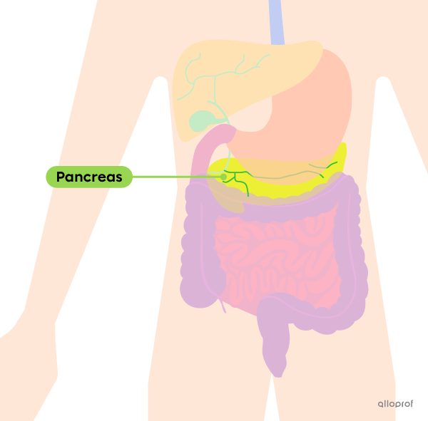
The pancreas is an elongated digestive gland, most of which is located behind the lower part of the stomach. It is an accessory gland to the digestive tract. The pancreatic juice produced by the pancreas is carried through a duct and discharged into the upper part of the small intestine. |
|
| Description | Function |
|---|---|
|
The intestinal glands are digestive glands integrated into the inner wall of the small intestine. They are located in cavities between the villi of the small intestine. They are glands integrated directly into the digestive tract. |
|

