There are over 100 000 different proteins in the human body. Each of them must be produced by the organism to fulfill a specific function.
Proteins
Protein Synthesis
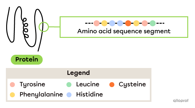
Proteins are macromolecules (large molecules) made up of a chain of amino acids of varying length.
There are 20 types of amino acids. These amino acids are small compounds that bind together to form a chain that can be short or very long. Once the protein is formed, the interactions between the amino acids force the chain to fold, giving each protein a distinctive shape.

Food is a source of protein. The digestive system allows the proteins to be digested by breaking the bonds that join the amino acids. The amino acids can then be absorbed into the bloodstream and distributed to the cells so they can synthesize new proteins.
Proteins are essential molecules for living organisms and viruses. Their functions vary greatly and are determined by their composition and three-dimensional shape.
-
Antibodies are proteins that recognize foreign bodies in order to trigger the body’s immune response.
-
Lactase is also a protein. It acts as an enzyme by breaking down lactose, which is a type of sugar found in many dairy products. Lactase is therefore involved in the chemical digestion of food.
-
Hemoglobin is a protein that binds and transports oxygen in the bloodstream.
-
Collagen is a protein with several functions, such as maintaining the cohesion and strength of the skin. Collagen is also present in other body tissues.
In order for the human body to function properly, cells must carry out chemical reactions, defend against attacks from foreign bodies, transport particles and more. Proteins have an important part to play in all these functions. The body must synthesize a wide variety of proteins to perform a wide variety of functions.
Protein synthesis consists of binding simple particles, called amino acids, to create a complex chain called a protein.
Protein synthesis is divided into two stages: transcription and translation.
These two processes are explained below.
In the cell nucleus (transcription)
-
The DNA is like a big cookbook. It contains all the information to synthesize all the proteins necessary for the human body. However, if only one protein is to be synthesized, only one recipe is required.
-
An enzyme reads the DNA and copies the unique recipe required. The copy of this recipe comes in the form of a molecule: the messenger RNA (mRNA).
Outside the cell nucleus (translation)
-
The mRNA leaves the nucleus and moves into the cytoplasm where protein synthesis takes place.
-
The ribosome acts as a cook. It reads the mRNA as a recipe and uses amino acids as ingredients to manufacture the desired protein.
-
Once protein synthesis is complete, it is released into the cytoplasm of the cell.
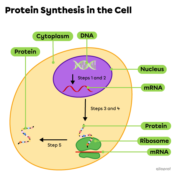
Transcription is the first step in protein synthesis. It consists of copying the genetic information included on a segment of the DNA by producing a messenger RNA (mRNA) molecule.
The information required to synthesize all the proteins in the body is contained in the DNA. Since the DNA is long and bulky, it cannot leave the cell nucleus to participate directly in protein synthesis. A smaller molecule capable of leaving the nucleus and transporting genetic information must be produced: the messenger ribonucleic acid, or mRNA.
There are some similarities between DNA and RNA molecules. For example, they both consist of sugars, nitrogenous bases and phosphate groups. The differences between these molecules are summarized in the following table.
| DNA | RNA | |
|---|---|---|
| Full name | Deoxyribonucleic acid | Ribonucleic acid |
| Type of sugar | Deoxyribose | Ribose |
| Types of nitrogenous bases | Adenine Thymine Guanine Cytosine |
Adenine Uracil Guanine Cytosine |
| Number of strands | Typically two strands | Typically one strand |
DNA transcription into mRNA takes place according to the following steps.
Initially, the DNA is made up of two strands with complementary nitrogenous bases (A-T pair and G-C pair). Free nitrogenous bases are also found scattered in the nucleus.
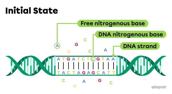
In the first step of transcription, an enzyme separates the two DNA strands. In addition, by using the free nitrogenous bases available in the nucleus, the enzyme matches each nitrogenous base in the DNA strand with a complementary nitrogenous base. This is how a strand of mRNA is formed.
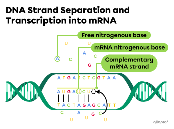
In the second and final step, the mRNA strand is completed. It dissociates from the DNA strand and may, if necessary, undergo a number of final modifications. It can then move outside the nucleus, into the cytoplasm of the cell.

The mRNA is therefore a complementary molecule to the DNA. During the formation of mRNA, the nitrogenous bases match in the same way as between two DNA strands. However, during mRNA synthesis, thymine (T) is replaced with uracil (U). The following table compares the pairing of nitrogenous bases between two DNA strands and during mRNA formation.
| Pairing of nitrogenous bases between two DNA strands (DNA strand-DNA strand) |
Nitrogenous base pairing during mRNA formation (DNA strand-mRNA strand) |
|---|---|
| Guanine-Cytosine Cytosine-Guanine Thymine-Adenine Adenine-Thymine |
Guanine-Cytosine Cytosine-Guanine Thymine-Adenine Adenine-Uracil |
Here is a strand of DNA.

What is the mRNA sequence corresponding to this DNA strand?
The DNA strand guiding the mRNA formation has the following sequence: TACGATGCA.
Based on the previous table, it is known that:
-
thymine (T) in DNA pairs with adenine (A) in mRNA
-
adenine (A) in DNA pairs with uracil (U) in mRNA
-
cytosine (C) in DNA pairs with guanine (G) in mRNA
-
guanine (G) in DNA pairs with cytosine (C) in mRNA
Therefore, the following mRNA strand is obtained.
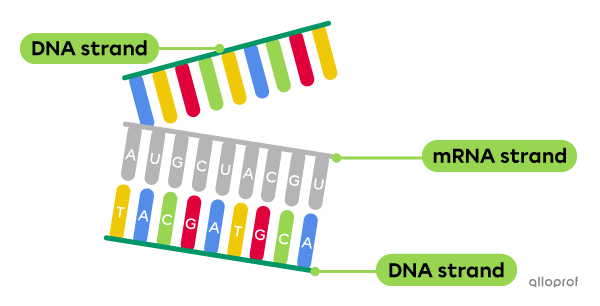
The sequence of this mRNA strand is therefore AUGCUACGU.
Now that the transcription step is complete, the cell can translate the messenger RNA (mRNA) into a protein.
-
Translation is the second step in protein synthesis. The cell’s ribosomes read the mRNA and synthesize the protein.
-
Transfer RNA (tRNA) is a type of RNA that binds to mRNA. It carries the amino acids that will form the protein.
-
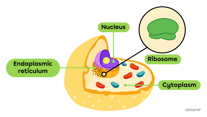
Ribosomes are organelles that are found within the cell on the surface of the endoplasmic reticulum.
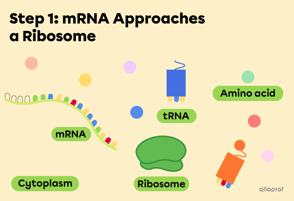
Step 1: When mRNA leaves the nucleus on the way to the cytoplasm, it approaches a ribosome. During this first step, several particles are already floating in the cytoplasm, including transfer RNA (tRNA) and amino acids.
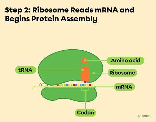
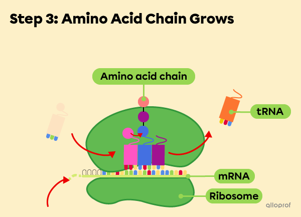
Step 2: The mRNA binds to the ribosome. The ribosome begins to read the mRNA in groups of three nitrogenous bases. Each nitrogenous base triplet is called a codon.
When the mRNA codon is read by the ribosome, a corresponding tRNA appears. The tRNA operates like a shuttle that transports a specific amino acid that will be part of the future protein.
Step 3: The ribosome continues to bind amino acids, creating a chain that grows longer.
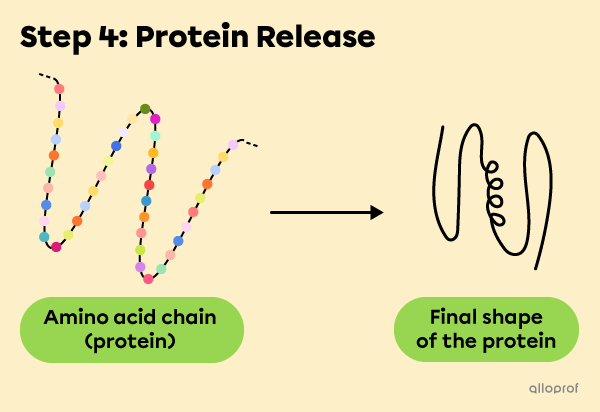
Step 4: Protein synthesis is complete and the amino acid chain is released into the cytoplasm. It folds and becomes a protein in its final three-dimensional shape.
An mRNA codon corresponds to a specific amino acid. For example, when an mRNA codon with nitrogenous bases U, U and C is read, an amino acid called phenylalanine is included in the protein chain. In other words, a UUC codon codes for phenylalanine.
The following reference table helps determine the codons responsible for the addition of a specific amino acid.
| 1st nitrogenous base | 2nd nitrogenous base | 3rd nitrogenous base | |||||||
| U | C | A | G | ||||||
| U | UUU | Phenylalanine | UCU | Serine | UAU | Tyrosine | UGU | Cysteine | U |
| UUC | UCC | UAC | UGC | C | |||||
| UUA | Leucine | UCA | UAA | - | UGA | - | A | ||
| UUG | UCG | UAG | UGG | Tryptophan | G | ||||
| C | CUU | CCU | Proline | CAU | Histidine | CGU | Arginine | U | |
| CUC | CCC | CAC | CGC | C | |||||
| CUA | CCA | CAA | Glutamine | CGA | A | ||||
| CUG | CCG | CAG | CGG | G | |||||
| A | AUU | Isoleucine | ACU | Threonine | AAU | Asparagine | AGU | Serine | U |
| AUC | ACC | AAC | AGC | C | |||||
| AUA | ACA | AAA | Lysine | AGA | Arginine | A | |||
| AUG | Methionine | ACG | AAG | AGG | G | ||||
| G | GUU | Valine | GCU | Alanine | GAU | Aspartic acid | GGU | Glycine | U |
| GUC | GCC | GAC | GGC | C | |||||
| GUA | GCA | GAA | Glutamic acid | GGA | A | ||||
| GUG | GCG | GAG | GGG | G | |||||
*The AUG triplet codes for methionine, but is also an initiation codon, signalling the beginning of the codon sequence.
**The UAA, UAG and UGA triplets do not code for amino acids but are end-of-translation codons, signalling the end of the codon sequence.
The amino acid sequence segment containing tyrosine-phenylalanine-histidine-cysteine-phenylalanine-tyrosine-leucine can be produced when the mRNA strand includes the codon sequence UAU-UUC-CAU-UGU-UUC-UAC-CUG.
