The female reproductive system consists of female reproductive organs, also referred to as female genitalia or female genital organs, involved in reproduction. It includes internal reproductive organs, located inside the body, and external reproductive organs, located mostly outside the body.
Internal reproductive organs, also referred to as internal genital organs, are involved in the ovarian and menstrual cycles and pregnancy. They are responsible for the production of ova and for gestation culminating in childbirth.
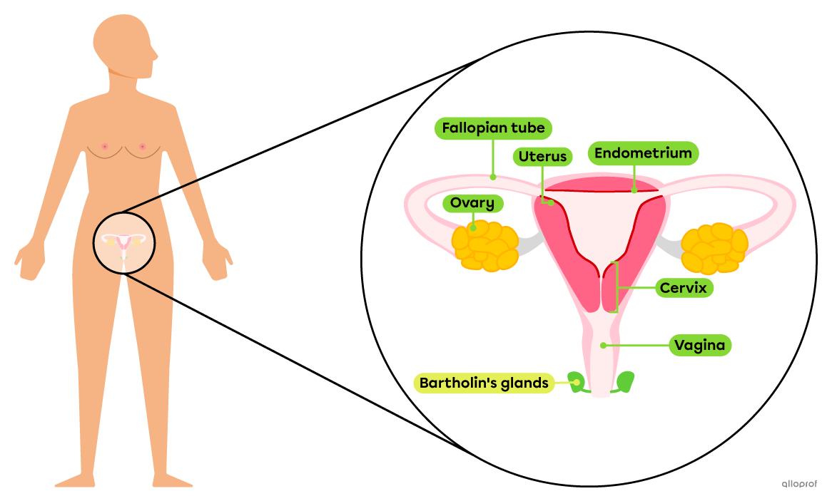
Note: The Bartholin's glands are located inside the body and open into the vulva.
The female reproductive system contains two ovaries. They are oval glands held in place by ovarian ligaments attached to the uterus. Each ovary is located next to the funnel-shaped opening of the Fallopian tube, without being attached to it.
The ovaries are the primary location for oogenesis, which is the production of female gametes. The ovaries also secrete hormones (estrogen and progesterone) that participate in the ovarian and menstrual cycles and play a role in physical changes during puberty in females.
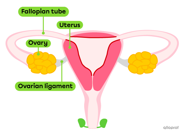
Fallopian tubes are two ducts, reaching the ovaries. They create a canal leading to the uterus. When an oocyte is released by the ovary, it is caught by the closest Fallopian tube. The oocyte is moved along the tube by the cilia covering its internal lining.
Fertilization occurs in the Fallopian tube. An oocyte becomes an ovum, then a zygote, which continues its path to the uterus. If there is no fertilization, the unfertilized oocyte simply moves through the Fallopian tube towards the uterus to be eventually evacuated with menstruation.
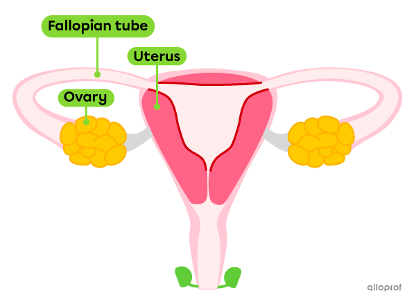
The uterus, commonly referred to as the womb, is the hollow, pear-shaped organ where the baby develops during pregnancy. In adult females, it is normally approximately 7 cm long and 5 cm wide[1], but stretches considerably during pregnancy.
The uterine walls are made of muscle tissue. These muscles contract to evacuate menstruation, which can sometimes be painful. This is called menstrual cramps. The muscles of the uterus also contract to deliver the baby and placenta during childbirth.
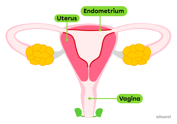
The inner lining of the uterus is called the endometrium.
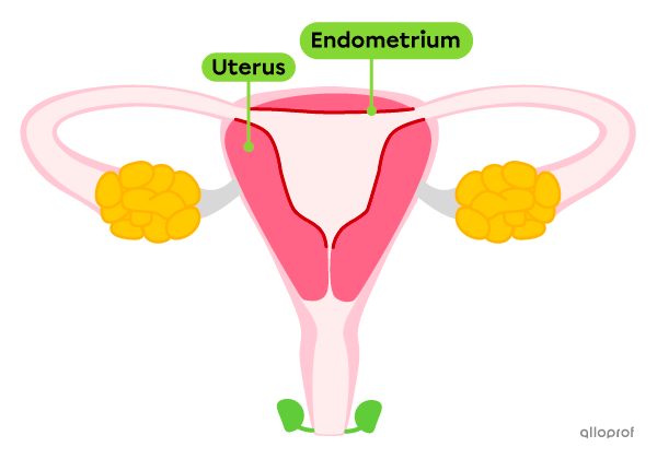
The inner cavity of the uterus is lined with a tissue rich in blood vessels, called the endometrium. The endometrium is required for the implantation of the embryo. It thickens during the menstrual cycle. If there is no fertilization, its superficial layer disintegrates and flows out through the vagina, a process known as menstruation.
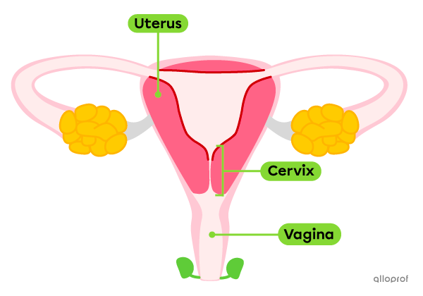
The cervix is the lowest part of the uterus, bordering the vagina. It keeps the developing baby inside the uterus during pregnancy, but it thins and widens its opening to let the baby through during birth. It also allows the flow of menstruation from the uterus to the vagina and the passage of spermatozoa into the uterus.
The vagina is a canal that connects the uterus to the outside of the body. Its entrance is approximately in the center of the vulva. The vagina is very stretchy. During sexual intercourse, it can receive the penis and the sperm. It also allows the passage of the baby during childbirth.
The vagina secretes lubricating fluids. It is also colonized by helpful bacteria that make up the vaginal flora. The vaginal flora forms a protective barrier against certain infections.
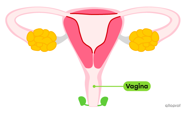
The word vagina is often used incorrectly to refer to the external genitalia. However, the vagina refers to an internal cavity. The word vulva should be used to refer to the external genitalia.
This video[2] briefly explains the history of gaps in the understanding of female reproductive health and their consequences. It is a subject that has long been sidelined by science, which has led to repercussions for women's physical and mental health.
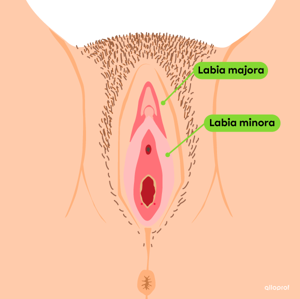
Together, the female external reproductive organs, also called external genital organs, form the vulva. It surrounds the entrance to the vagina and includes the labia minora and labia majora, clitoral hood, glans clitoris, hymen and Bartholin's glands.
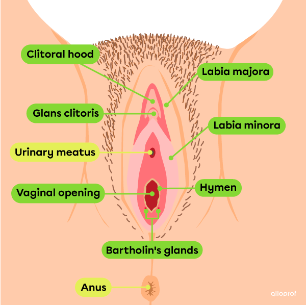
Note: The urinary meatus is part of the urinary system and the anus is part of the digestive system.
The labia, sometimes referred to as the lips, is a set of skin folds.
The labia minora, sometimes called the inner lips, is a set of thin folds surrounding the urinary meatus and the vaginal opening. The function of the labia minora is to protect these structures.
The labia majora, sometimes called the outer lips, is a set of thicker skin folds, covered with hair. They surround the labia minora and protect them.
The appearance of the labia majora and labia minora varies greatly from person to person.
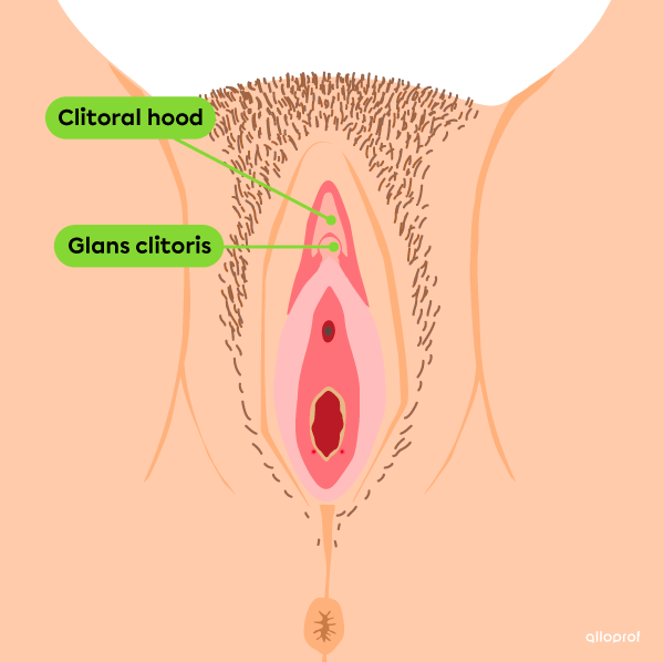
The clitoris is an organ found both outside and inside the body. From the outside, the clioral hood and the glans clitoris are visible. The clitoral hood is a skin fold that covers the glans clitoris. Most of the clitoris is hidden and extends on either side of the vagina inside the body.
The clitoris is made up of erectile tissue and is very sensitive to touch. It fills with blood and increases in size when excited. Its role is to provide sexual pleasure.
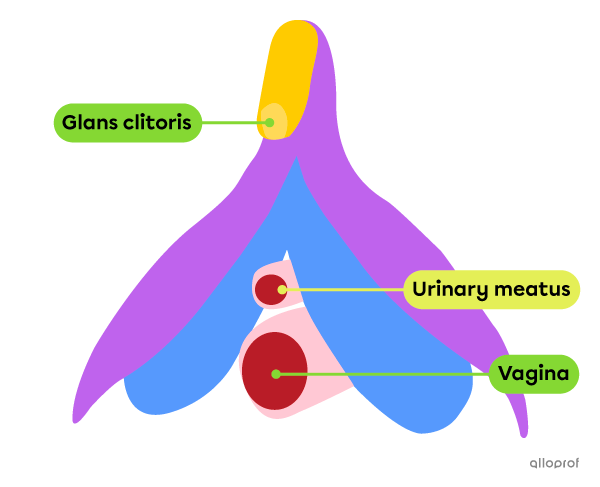
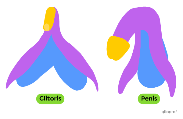
The genital tubercle is a structure present in both male and female fetuses. The tissues of this tubercle differentiate during the first months of pregnancy to form either female or male genitalia. The clitoris and the penis are made of the same tissues. They develop differently, but still have a lot in common. In addition to being shaped quite similarly, they are both erectile organs, sensitive to touch.
The clitoris was first identified in ancient times. Back then, only a small part of its anatomy was known. It wasn't until the 20th century (over 2 000 years later!) that its full anatomy was discovered. Watch this fun short documentary[3] to learn more about the anatomy and history of this organ.
The hymen is a half-moon or crown shaped membrane that usually partially covers the vaginal opening. The appearance, thickness, and coverage of the hymen varies greatly from person to person. During some physical activities or vaginal penetration, such as sexual intercourse or the application of a tampon, the hymen can stretch. Since the flexibility of the hymen varies, it can tear in some cases. The tear may or may not be accompanied by bleeding.
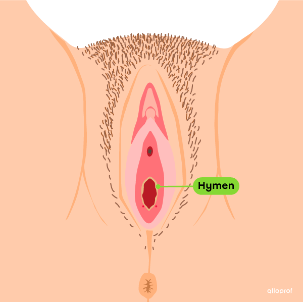
For a long time, it was believed that an intact hymen was evidence that vaginal penetration has never occurred. However, we now know that not only can the hymen stretch and remain intact after penetration, it can also rupture without penetration taking place. Therefore, the appearance of the hymen cannot be used to confirm whether vaginal penetration has ever occurred.
The Bartholin’s glands, also called the greater vestibular glands, are located inside the body on either side of the vaginal opening. The glands open into the vulva, where they secrete a lubricating fluid.

1. Canadian Cancer Society. (s.d.). The uterus. https://cancer.ca/fr/cancer-information/cancer-types/uterine/what-is-ut…
2. Seeker. (2021, June 29). Why We Know So Little About Women’s Bodies [Video]. https://www.seeker.com/videos/health/why-we-know-so-little-about-womens…
3. Malépart-Traversy, L. (2016). Le clitoris [documentaire animé]. Graduation film, Mel Hoppenheim School of Cinema, Concordia University (Montréal, Canada).